Home » Without Label » Bones In Leg Diagram - Thumb Elbow Bone Human Leg Anatomy Others Angle Hand Anatomy Png Pngwing / The tibia and fibula form the ankle joint with the talus,.
Bones In Leg Diagram - Thumb Elbow Bone Human Leg Anatomy Others Angle Hand Anatomy Png Pngwing / The tibia and fibula form the ankle joint with the talus,.
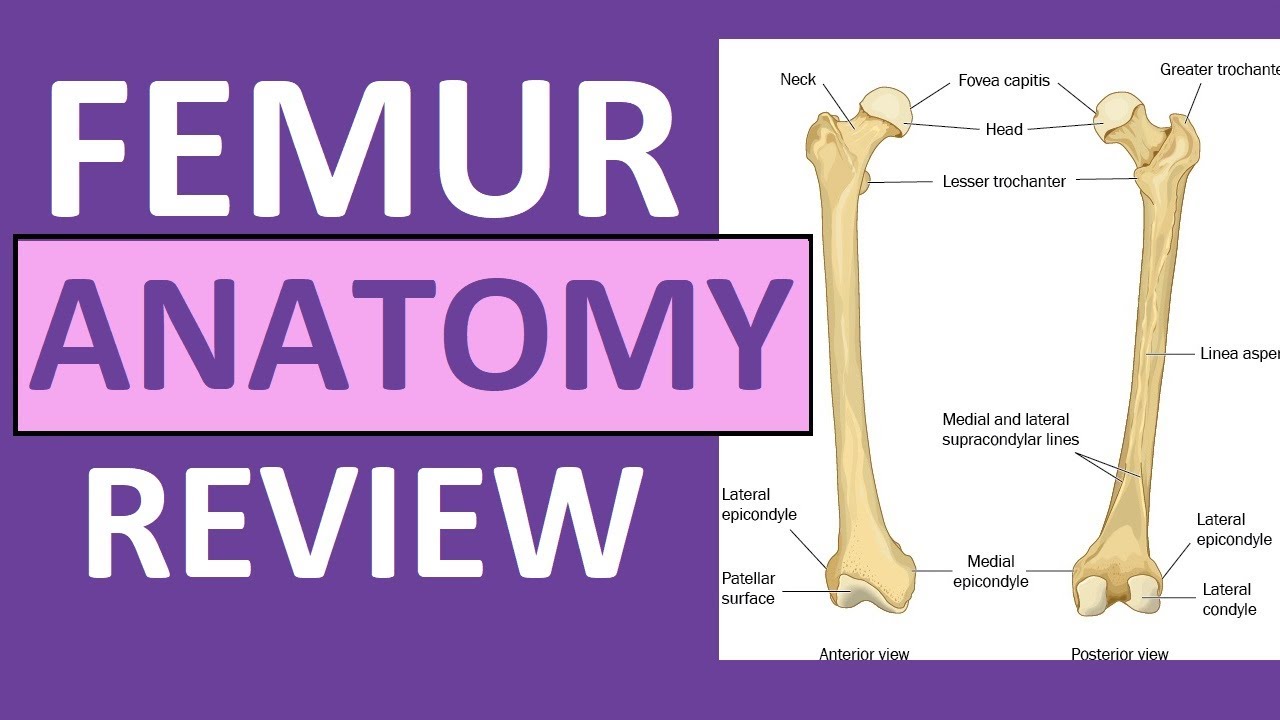
Bones In Leg Diagram - Thumb Elbow Bone Human Leg Anatomy Others Angle Hand Anatomy Png Pngwing / The tibia and fibula form the ankle joint with the talus,.. The thigh bone, or femur, is the large upper leg bone that connects the lower leg bones (knee joint) to the pelvic bone (hip joint). The smaller lateral bone of the lower leg. This allows weight to be distributed either anteriorly or posteriorly throughout the foot. The tarsal bones in the foot are located amongst tibia, metatarsal bones, and fibula. Its lower end helps create the knee joint.
The smaller lateral bone of the lower leg. The femur, or thighbone, is the longest and largest bone in the human body. Human knee anatomy diagram free vector. The tibia and fibula are two long bones that run parallel to each other, forming the scaffold of the leg and providing attachment points for many muscles. The authors explore how digitizing one of the seven basic quality tools—the fishbone diagram—using mind mapping can significantly improve the tool.
Tibia And Fibula Anatomy Of Leg Bones Anatomy Physiology Youtube from i.ytimg.com The femur is the single bone of the thigh. Health diagram bone skeleton leg knee science anchor chart human human body. Bone of pelvis pics 12 photos of the bone of pelvis pics , bone. Bone test anatomy and physiology 12 photos of the bone test anatomy and physiology anatomy and physiology bone lab test, anatomy and physiology bone markings test, anatomy and physiology bone practical test, anatomy and physiology bone tissue test, anatomy and physiology test on bone tissue, bone, anatomy and. The hip itself is a ball and socket joint, much like the shoulder.the structures necessary to create this joint are the socket, the joint capsule, muscle, ligaments, and the neck. These are the femur, patella, tibia, fibula, tarsal bones, metatarsal bones, and phalanges (see figure 6.51). The tibia and fibula are the bones of the lower leg. The talocrual joint is made up of three main bones.
The patella is the kneecap and articulates with the distal femur.
Human knee anatomy diagram free vector. Bone test anatomy and physiology 12 photos of the bone test anatomy and physiology anatomy and physiology bone lab test, anatomy and physiology bone markings test, anatomy and physiology bone practical test, anatomy and physiology bone tissue test, anatomy and physiology test on bone tissue, bone, anatomy and. Browse 7,035 leg bone stock photos and images available, or search for leg bone xray or human leg bone to find more great stock photos and pictures. The thigh bone, or femur, is the large upper leg bone that connects the lower leg bones (knee joint) to the pelvic bone (hip joint). The tibia is much larger than the fibula and bears almost all of the body's weight. Related posts of diagram of leg bones bone of pelvis pics. Leg pain can also be caused by blood clots, varicose veins or poor circulation. These muscles work together to produce movements such as standing, walking, running, and jumping. The lower leg is comprised of two bones, the tibia and the smaller fibula. The diagram of bones in the ankle and foot is given below: The smaller lateral bone of the lower leg. The talocrual joint is made up of three main bones. The proximal portion of the tibia is tibial plateau which acts as a cusp for the knee, the distal portion tapers into the medial malleoli and the concave surface which articulates with the talus at the ankle joint.
Ankle & lower leg anatomy. At the same time, the bones and joints of the leg and foot must be strong enough to support the body's weight while remaining. The lower leg is comprised of two bones, the tibia and the smaller fibula. The tarsal bones in the foot are located amongst tibia, metatarsal bones, and fibula. The knee joint is the largest joint in the body and is primarily a hinge joint, although some sliding and rotation occur.
Leg Fracture Child from www.fairview.org Also called the shin bone, the tibia is the longer of the two bones in the. The talocrual joint is made up of three main bones. Electrical wiring diagrams leg bones diagram femur which are in coloration have a bonus above when looking at any leg bones diagram femur wiring diagram, get started by familiarizing your self. The medial, larger bone of the lower leg. The tarsal bones in the foot are located amongst tibia, metatarsal bones, and fibula. This allows weight to be distributed either anteriorly or posteriorly throughout the foot. Another bone that is part of the lower leg and the knee joint is called the fibula.this is a bone located on the lateral, or outer part, of the lower leg and is more commonly known as the calf bone. Ankle & lower leg anatomy.
Some common causes of leg pain include:
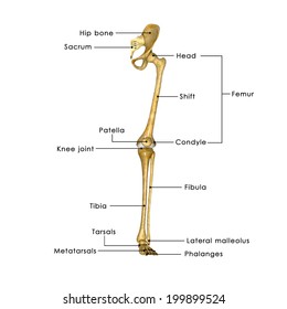
The bones together make up the hip. Browse 7,035 leg bone stock photos and images available, or search for leg bone xray or human leg bone to find more great stock photos and pictures. The smaller lateral bone of the lower leg. Human knee anatomy diagram free vector. The tibia is much larger than the fibula and bears almost all of the body's weight. To explain the term in layman's language, it is the heel bone in the skeletal system. Bone diagram forehead (frontal bone) nose bones (nasals) cheek bone (zygoma) upper jaw (maxilla) lower jaw (mandible) breast bone (sternum) upper arm bone (humerus) lower arm bone (ulna) thigh bone (femur) collar bone (clavicle) toe bones (phalanges) ankle bones (tarsals) kneecap (patella) shin bone 10 october 2007 (original upload date) These muscles work together to produce movements such as standing, walking, running, and jumping. Some types of leg pain can be traced to problems in your lower spine. The lower leg is comprised of two bones, the tibia and the smaller fibula. At the same time, the bones and joints of the leg and foot must be strong enough to support the body's weight while remaining. The bones of the hip include the femur, the ilium, the ischium, and the pubis.
Bone on side of the foot Health diagram bone skeleton leg knee science anchor chart human human body. The proximal portion of the tibia is tibial plateau which acts as a cusp for the knee, the distal portion tapers into the medial malleoli and the concave surface which articulates with the talus at the ankle joint. The major bones of the leg are the femur (thigh bone), tibia (shin bone), and adjacent fibula, and these are all long bones.the patella (kneecap) is the sesamoid bone in front of the knee.most of the leg skeleton has bony prominences and margins that can be palpated and some serve as anatomical landmarks that define the extent of the leg. Fish(bone) stories (quality progress) the method behind the fishbone diagram is older than many of its users.
Labelled Bones Leg High Res Stock Images Shutterstock from image.shutterstock.com These are the femur, patella, tibia, fibula, tarsal bones, metatarsal bones, and phalanges (see figure 6.51). The thigh bone, or femur, is the large upper leg bone that connects the lower leg bones (knee joint) to the pelvic bone (hip joint). Health diagram bone skeleton leg knee science anchor chart human human body. The hip itself is a ball and socket. The tibia and fibula are two long bones that run parallel to each other, forming the scaffold of the leg and providing attachment points for many muscles. Related posts of bones leg diagram picture bone test anatomy and physiology. Blank leg bones diagram : The fibula is mainly a muscle attachment point and is used to help maintain balance.
The medial, larger bone of the lower leg.
The proximal portion of the tibia is tibial plateau which acts as a cusp for the knee, the distal portion tapers into the medial malleoli and the concave surface which articulates with the talus at the ankle joint. The tibia and fibula are two long bones that run parallel to each other, forming the scaffold of the leg and providing attachment points for many muscles. The knee joint is the largest joint in the body and is primarily a hinge joint, although some sliding and rotation occur. The talus the weight of your body is transferred from the tiba to the talus. The major bones of the leg are the femur (thigh bone), tibia (shin bone), and adjacent fibula, and these are all long bones.the patella (kneecap) is the sesamoid bone in front of the knee.most of the leg skeleton has bony prominences and margins that can be palpated and some serve as anatomical landmarks that define the extent of the leg. The tibia and the fibula, at the top of the ankle joint. The femur is the single bone of the thigh. These muscles work together to produce movements such as standing, walking, running, and jumping. The tibia, commonly known as the 'shin bone', is the largest and most medial of the two.you can palpate its anterior border when you run your finger down the anterior aspect of your leg. The bones of the leg are the femur, tibia, fibula and patella. Browse 7,035 leg bone stock photos and images available, or search for leg bone xray or human leg bone to find more great stock photos and pictures. The bones together make up the hip. Its lower end helps create the knee joint.